Home

postgraduálna škola Inak V reakcii na lps staining výkaz štyri nastaviť

Cells | Free Full-Text | LPS-Induced Systemic Inflammation Affects the  Dynamic Interactions of Astrocytes and Microglia with the Vasculature of  the Mouse Brain Cortex
Cells | Free Full-Text | LPS-Induced Systemic Inflammation Affects the Dynamic Interactions of Astrocytes and Microglia with the Vasculature of the Mouse Brain Cortex

Metformin decreases LPS-induced inflammatory response in rabbit annulus  fibrosus stem/progenitor cells by blocking HMGB1 release | Aging
Metformin decreases LPS-induced inflammatory response in rabbit annulus fibrosus stem/progenitor cells by blocking HMGB1 release | Aging

Neutrophil extracellular traps are indirectly triggered by  lipopolysaccharide and contribute to acute lung injury | Scientific Reports
Neutrophil extracellular traps are indirectly triggered by lipopolysaccharide and contribute to acute lung injury | Scientific Reports

Ginsenoside Rg1 attenuates LPS-induced chronic renal injury by inhibiting  NOX4-NLRP3 signaling in mice - ScienceDirect
Ginsenoside Rg1 attenuates LPS-induced chronic renal injury by inhibiting NOX4-NLRP3 signaling in mice - ScienceDirect
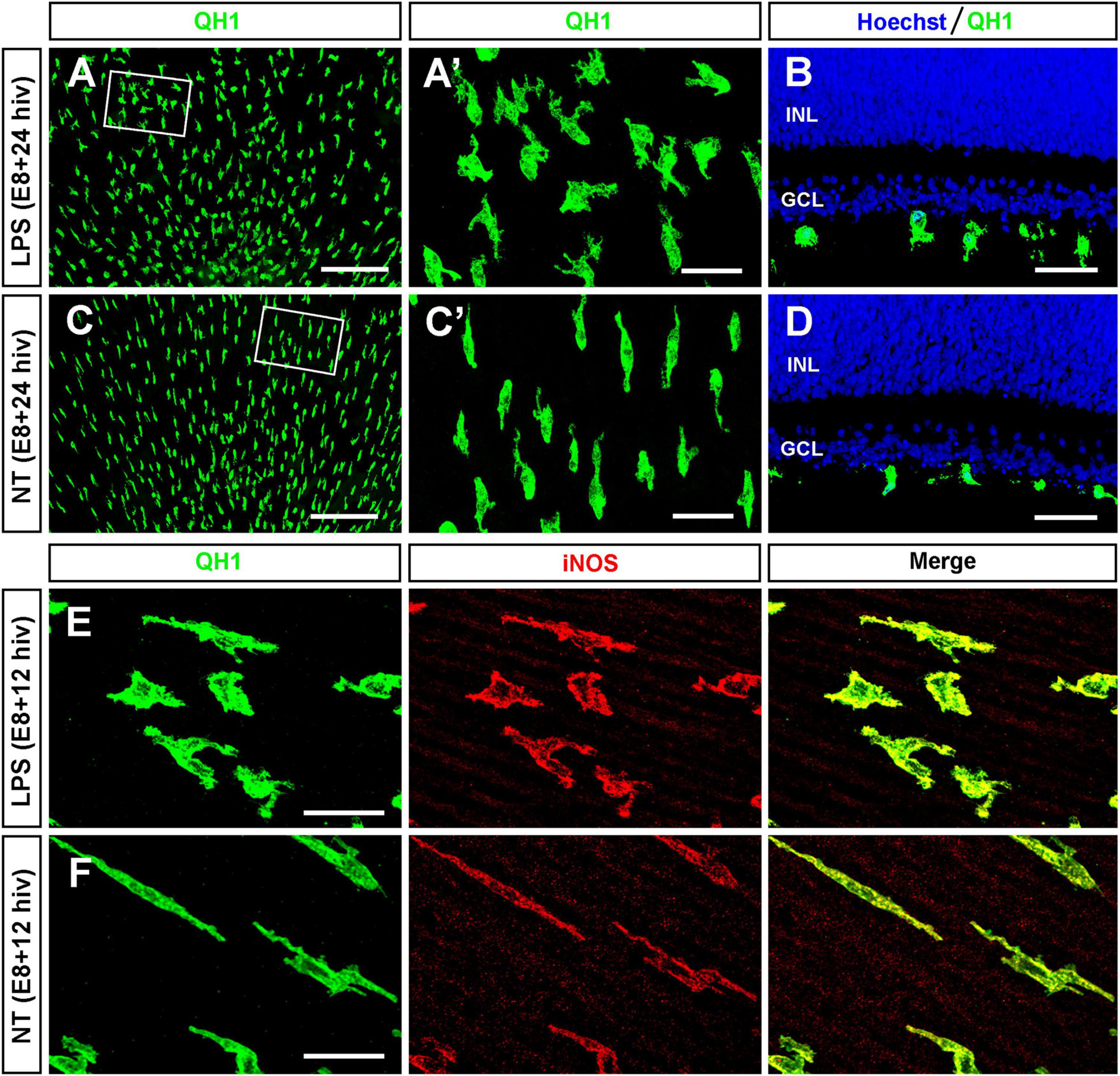
Silver stained SDS-PAGE analysis of LPS extracted from E. coli O104 strains  and resistant mutants.
Silver stained SDS-PAGE analysis of LPS extracted from E. coli O104 strains and resistant mutants.

Neuronal Mitochondria Modulation of LPS-Induced Neuroinflammation | Journal  of Neuroscience
Neuronal Mitochondria Modulation of LPS-Induced Neuroinflammation | Journal of Neuroscience

Lipopolysaccharides (LPS) Antibody (ABIN479062)

Frontiers | LPS-stimulated microglial cells promote ganglion cell death in  organotypic cultures of quail embryo retina
Frontiers | LPS-stimulated microglial cells promote ganglion cell death in organotypic cultures of quail embryo retina

Purification and Visualization of Lipopolysaccharide from Gram-negative  Bacteria by Hot Aqueous-phenol Extraction | Protocol
Purification and Visualization of Lipopolysaccharide from Gram-negative Bacteria by Hot Aqueous-phenol Extraction | Protocol
IJMS | Free Full-Text | Complete Characterization of the O-Antigen from the  LPS of Aeromonas bivalvium
IJMS | Free Full-Text | Complete Characterization of the O-Antigen from the LPS of Aeromonas bivalvium

Lipopolysaccharide (LPS) staining in the human superior temporal lobe... |  Download Scientific Diagram
Lipopolysaccharide (LPS) staining in the human superior temporal lobe... | Download Scientific Diagram

Analysis of lipopolysaccharide profiles by silver staining. Silver... |  Download Scientific Diagram
Analysis of lipopolysaccharide profiles by silver staining. Silver... | Download Scientific Diagram

LPS structure analysis. (A) Silver staining of purified LPS. Each lane... |  Download Scientific Diagram
LPS structure analysis. (A) Silver staining of purified LPS. Each lane... | Download Scientific Diagram

Metformin decreases LPS-induced inflammatory response in rabbit annulus  fibrosus stem/progenitor cells by blocking HMGB1 release - Figure f4 | Aging
Metformin decreases LPS-induced inflammatory response in rabbit annulus fibrosus stem/progenitor cells by blocking HMGB1 release - Figure f4 | Aging

Enriched LPS Staining within the Germinal Center of a Lymph Node from an  HIV-Infected Long-Term Nonprogressor but Not from Progressors
Enriched LPS Staining within the Germinal Center of a Lymph Node from an HIV-Infected Long-Term Nonprogressor but Not from Progressors
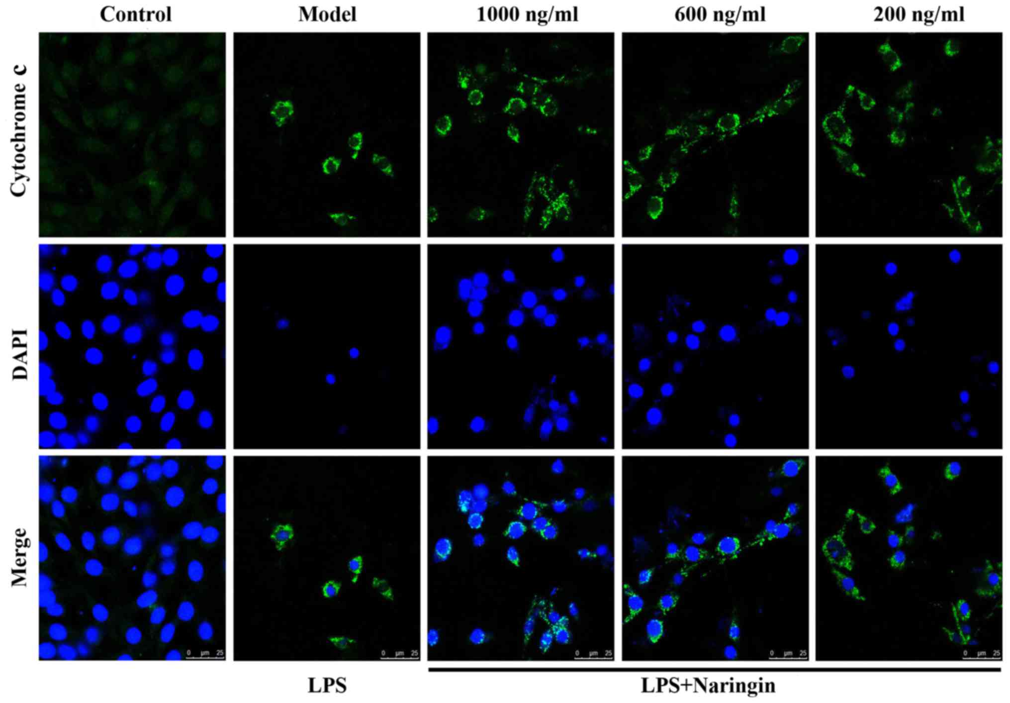
Protective effect of naringin against the LPS-induced apoptosis of PC12  cells: Implications for the treatment of neurodegenerative disorders
Protective effect of naringin against the LPS-induced apoptosis of PC12 cells: Implications for the treatment of neurodegenerative disorders
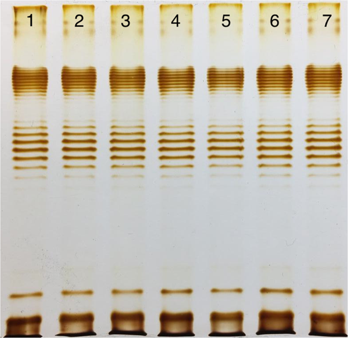
High-throughput LPS profiling as a tool for revealing of bacteriophage  infection strategies | Scientific Reports
High-throughput LPS profiling as a tool for revealing of bacteriophage infection strategies | Scientific Reports
Silver staining of LPS. LPS was separated on a 5 to 20% gradient... |  Download Scientific Diagram
Silver staining of LPS. LPS was separated on a 5 to 20% gradient... | Download Scientific Diagram

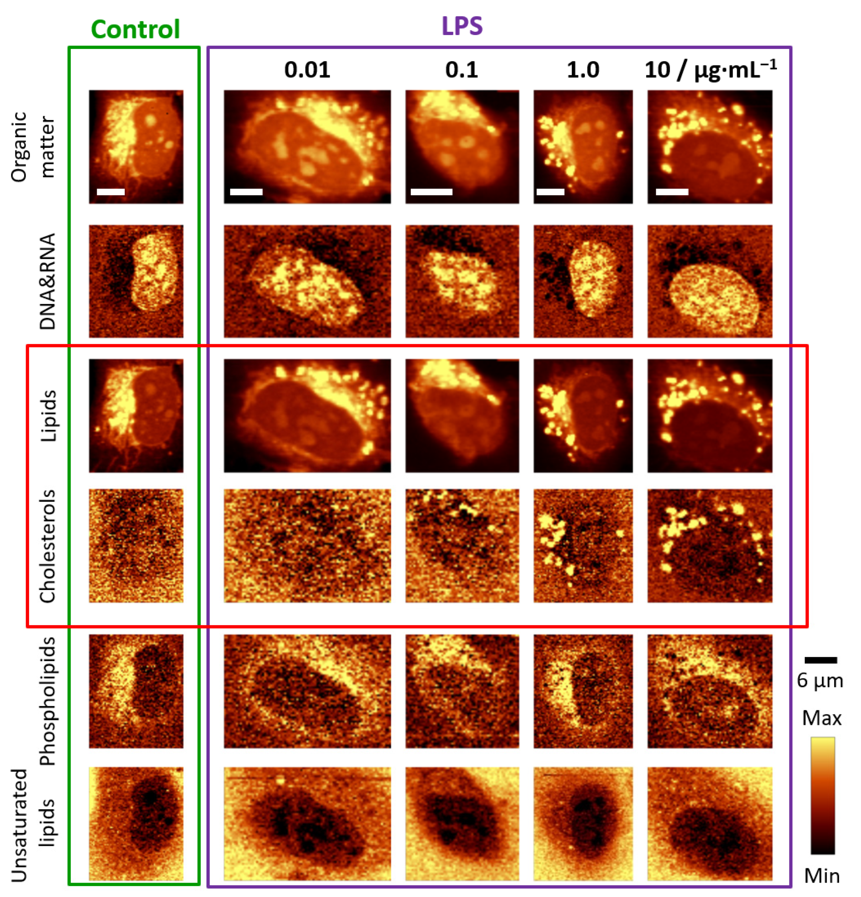
Cells | Free Full-Text | Lipid Droplets Formation Represents an Integral  Component of Endothelial Inflammation Induced by LPS
Cells | Free Full-Text | Lipid Droplets Formation Represents an Integral Component of Endothelial Inflammation Induced by LPS

JCI - Induction of B7-1 in podocytes is associated with nephrotic syndrome

Immunofluorescence staining of LPS-induced mTORC1 translocation to the... |  Download Scientific Diagram
Immunofluorescence staining of LPS-induced mTORC1 translocation to the... | Download Scientific Diagram
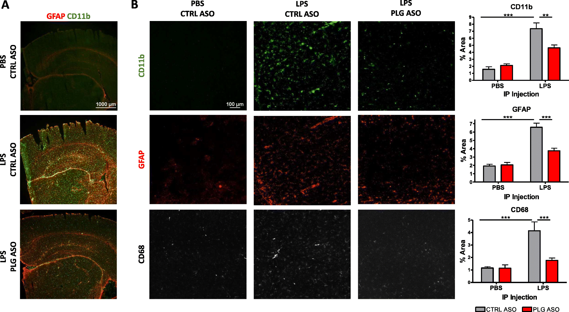
Plasminogen mediates communication between the peripheral and central  immune systems during systemic immune challenge with lipopolysaccharide |  Journal of Neuroinflammation | Full Text
Plasminogen mediates communication between the peripheral and central immune systems during systemic immune challenge with lipopolysaccharide | Journal of Neuroinflammation | Full Text
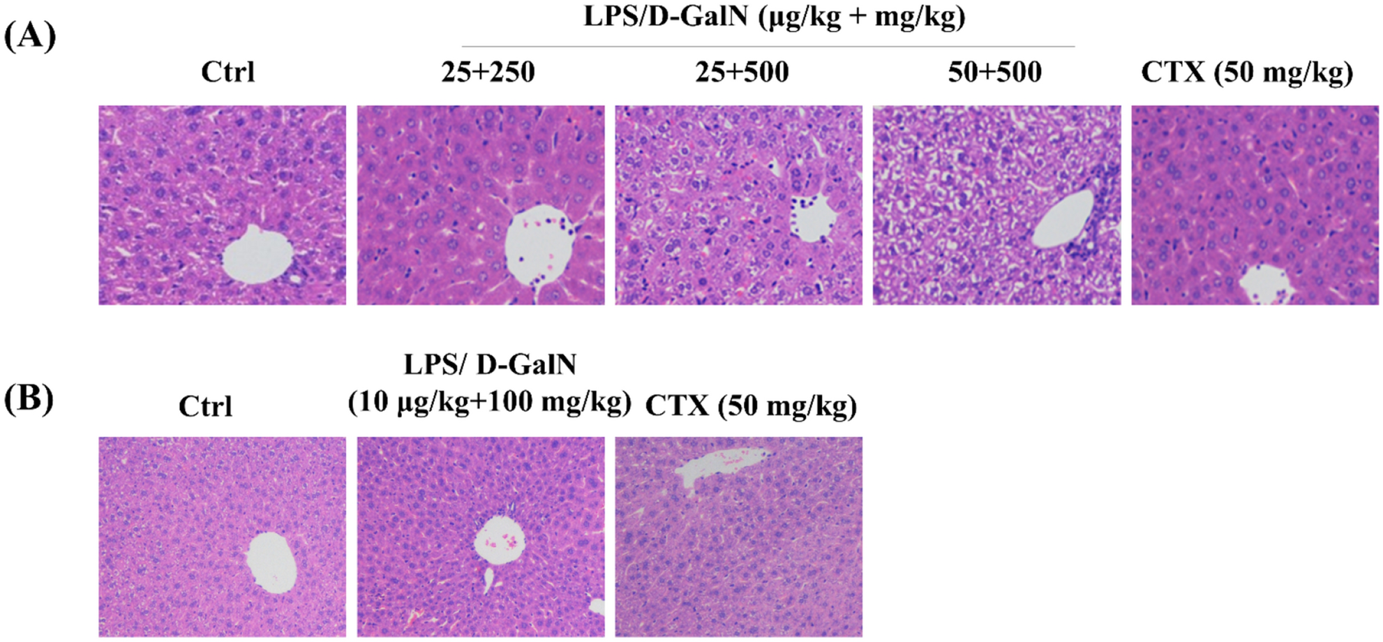
Co-administration of lipopolysaccharide and d-galactosamine induces  genotoxicity in mouse liver | Scientific Reports
Co-administration of lipopolysaccharide and d-galactosamine induces genotoxicity in mouse liver | Scientific Reports
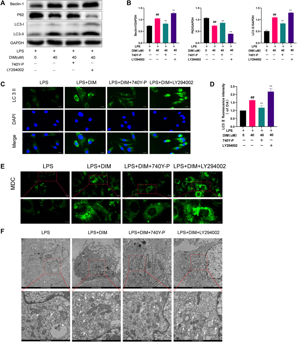
Lipid A LPS Polyclonal Antibody (PA1-73178)

Frontiers | 3,3′-diindolylmethane inhibits LPS-induced human chondrocytes  apoptosis and extracellular matrix degradation by activating  PI3K-Akt-mTOR-mediated autophagy
Frontiers | 3,3′-diindolylmethane inhibits LPS-induced human chondrocytes apoptosis and extracellular matrix degradation by activating PI3K-Akt-mTOR-mediated autophagy

A mutant Escherichia coli that attaches peptidoglycan to lipopolysaccharide  and displays cell wall on its surface | eLife
A mutant Escherichia coli that attaches peptidoglycan to lipopolysaccharide and displays cell wall on its surface | eLife

The Virtual Edge
The Virtual Edge